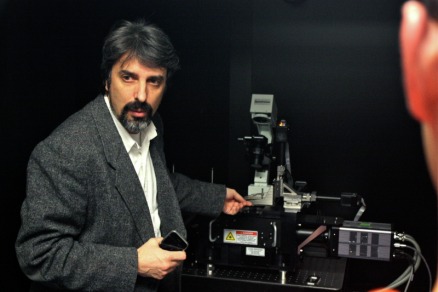
On a field trip to the Center for Biophotonics research center in Sacramento, we got to see a bunch of really interesting equipment. Above, one of the researchers shows us the highest-resolution optical microscope commercially available. It uses Moire patterns to focus down to 15 nanometers in size (that's 15 millionths of a millimeter).
This quarter, I am taking a particularly interesting class called 'Introduction to Biophotonics'. If you’ve never heard of Biophotonics, you’re in the same boat as everyone else I’ve told about it. I didn’t know what it was until I took the class, but I can testify that it is incredibly interesting. If I were pursuing a more science-oriented path, I would definitely consider a career in Biophotonics.
I suppose I should clarify what it is before I say any more about it. Biophotonics is essentially the combination of the fields of biology and photonics. If you’re as informed as the average layman, you’ll know that biology is the study of living organisms, but you won't know that photonics is the study of light (the real definition is slightly more complex, but we’ll keep it simple). Therefore, biophotonics is the study of how light can be used in biology and medicine.
I suppose I should clarify what it is before I say any more about it. Biophotonics is essentially the combination of the fields of biology and photonics. If you’re as informed as the average layman, you’ll know that biology is the study of living organisms, but you won't know that photonics is the study of light (the real definition is slightly more complex, but we’ll keep it simple). Therefore, biophotonics is the study of how light can be used in biology and medicine.
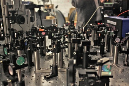
Another set up at the Center for Biophotonics. Each one of those little posts holds a prism, filter, or diffractor that manipulates the laser beam that passes through them.
Perhaps the best thing about biophotonics is that it is very non-invasive. Consider the following examples that show some of the applications of biophotonics.
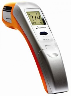
Infrared Thermometer:
The infrared thermometer uses infrared light to take your temperature. You just swipe it across your temple, and it instantly displays your temperature. It does this by measuring the amount of infrared radiation emitted by your temporal artery, which runs across your temple. Our bodies emit infrared radiation according to how much heat they produce. The hottest parts give off the most infrared radiation while the coolest parts give off the least. Our blood is a good measure of our core temperature, so measuring the infrared radiation given off by an artery (particularly one that’s close to the surface of our skin) is a good way to measure our temperature. No more waiting!
The infrared thermometer uses infrared light to take your temperature. You just swipe it across your temple, and it instantly displays your temperature. It does this by measuring the amount of infrared radiation emitted by your temporal artery, which runs across your temple. Our bodies emit infrared radiation according to how much heat they produce. The hottest parts give off the most infrared radiation while the coolest parts give off the least. Our blood is a good measure of our core temperature, so measuring the infrared radiation given off by an artery (particularly one that’s close to the surface of our skin) is a good way to measure our temperature. No more waiting!
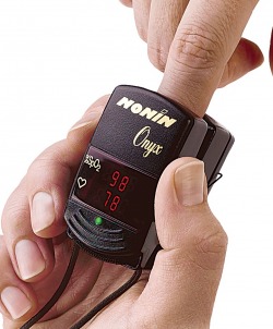
Pulse Oximeter:
This is a better example of how biophotonics is minimally invasive. In the days of old, there was no easy way to discover how much oxygen was being carried in a person’s blood. The only way was to take a blood sample from an artery and run time-consuming tests. This little device, however, makes all that obsolete.
Here’s what it does. You clamp it to the end of your finger, wait a few seconds, and then the LCD screen displays how oxygenated your blood is (typically around 98%). It’s that simple. How does it do it?
On one side of the clamp is a red light that shines through your finger to the other side. On the other side of the clamp, there is a detector that senses the color of the light passing through the finger. If you’ve ever shone a flashlight through your finger, you’ll know that the white light of the flashlight emerges on the other side of the finger as red light. If you know anything about the properties of light and color, you’ll know that things basically get their color by what wavelengths of light they absorb, and what wavelengths of light they reflect (if you’re not familiar with this, the quirky video below shows it pretty well). So for our flesh, red is reflected while the other colors are absorbed, chiefly because of blood and other tissues which take on a red color.
In school, you probably saw diagrams of our arteries and veins with arteries colored red and veins colored blue. Many people get confused and take this to mean that blood carrying oxygen (found in arteries) is colored red and blood lacking oxygen (found in veins) is colored blue. This is not the case. In reality, oxygenated blood is colored bright red and deoxygenated blood is colored dark red. In fact, the more oxygen blood is carrying, the brighter the color of red, and vice versa. You can now probably guess how the pulse oximeter works. It senses the exact color of the red coming through your finger and by that color, it can determine how much oxygen your blood is carrying.
This has many useful applications that would not be possible with the older, more time-consuming methods. The most obvious application is in emergency situations, where the oxygen content of a person’s blood might predict the success or failure of a particular course of action. There are less obvious applications too. Pilots at high altitudes can use them to determine if they are receiving enough oxygen from the air, or if they need to use on-board oxygen tanks (which is expensive, and best not used if it can be avoided). Even athletes can use them to track respiratory progress in training.
These tools are just a couple of applications of biophotonics. What I find most fascinating, however, are the applications on the cutting edge of research.
I could go in several directions at this point, but for the sake of brevity, I’ll highlight just one of the areas in which biophotonics is showing very exciting possibilities: cancer diagnosis.
Currently, there are few ways of conducting reliable cancer diagnostics that don’t involve invasive techniques such as biopsy. Often, this results in the patient getting a bunch of tissue cut out of them only to find out that they don’t have malignant cancer. Specifically for breast cancer, nearly 85% of all open surgical breast biopsies are found to be clear of cancer. In the U.S. alone, that’s nearly 19,000 women every week losing valuable tissue for no reason. Unsurprisingly, this process leaves many women feeling physical and emotional trauma.
Biophotonics shows promising signs, however, that such methods may not be necessary in the future. Now brace yourself for a little more science. I’ll keep it simple, both because I haven’t really learned the full complexity of it, and because most readers care more for the application than the theory.
So when light comes into contact with living material, it interacts. Specifically, when light hits a particular molecule, it will interact with the chemical bonds in the molecule and cause tiny changes in the characteristics of the light. It does this by momentarily absorbing a photon (a particle of light) at a time, which “excites” the molecule to a higher energy state. After a moment, the molecule “relaxes” and spits the photon back out, but the photon that comes out is slightly different than the one that went in. The molecule might emit a photon with slightly less energy than the photon that it absorbed (which would leave the molecule still in a slightly excited state). Or, conversely, the molecule might emit a photon with slightly more energy than the photon that it absorbed. This would leave the molecule more relaxed than it had been before absorbing the photon.
Particular types of molecules will exhibit different types of behaviors. Furthermore, any given molecule will interact differently with lights of different energies (effectively, different colors). So by systematically studying a molecule’s (or cell’s) interaction with different colors of light, that molecule’s chemical makeup can be determined. More importantly for cancer diagnosis, molecules and cells can be identified by their interactions with light. So a cancerous cell will interact with light differently than a healthy cell.
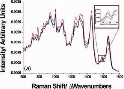
The visual representation of a molecule’s interaction with different colors of light is called a Raman Spectrum, shown below. As you can see, the spectrum of a healthy cell (black) is visibly different from the spectrum of a cancerous cell (red).
This has many useful applications that would not be possible with the older, more time-consuming methods. The most obvious application is in emergency situations, where the oxygen content of a person’s blood might predict the success or failure of a particular course of action. There are less obvious applications too. Pilots at high altitudes can use them to determine if they are receiving enough oxygen from the air, or if they need to use on-board oxygen tanks (which is expensive, and best not used if it can be avoided). Even athletes can use them to track respiratory progress in training.
These tools are just a couple of applications of biophotonics. What I find most fascinating, however, are the applications on the cutting edge of research.
I could go in several directions at this point, but for the sake of brevity, I’ll highlight just one of the areas in which biophotonics is showing very exciting possibilities: cancer diagnosis.
Currently, there are few ways of conducting reliable cancer diagnostics that don’t involve invasive techniques such as biopsy. Often, this results in the patient getting a bunch of tissue cut out of them only to find out that they don’t have malignant cancer. Specifically for breast cancer, nearly 85% of all open surgical breast biopsies are found to be clear of cancer. In the U.S. alone, that’s nearly 19,000 women every week losing valuable tissue for no reason. Unsurprisingly, this process leaves many women feeling physical and emotional trauma.
Biophotonics shows promising signs, however, that such methods may not be necessary in the future. Now brace yourself for a little more science. I’ll keep it simple, both because I haven’t really learned the full complexity of it, and because most readers care more for the application than the theory.
So when light comes into contact with living material, it interacts. Specifically, when light hits a particular molecule, it will interact with the chemical bonds in the molecule and cause tiny changes in the characteristics of the light. It does this by momentarily absorbing a photon (a particle of light) at a time, which “excites” the molecule to a higher energy state. After a moment, the molecule “relaxes” and spits the photon back out, but the photon that comes out is slightly different than the one that went in. The molecule might emit a photon with slightly less energy than the photon that it absorbed (which would leave the molecule still in a slightly excited state). Or, conversely, the molecule might emit a photon with slightly more energy than the photon that it absorbed. This would leave the molecule more relaxed than it had been before absorbing the photon.
Particular types of molecules will exhibit different types of behaviors. Furthermore, any given molecule will interact differently with lights of different energies (effectively, different colors). So by systematically studying a molecule’s (or cell’s) interaction with different colors of light, that molecule’s chemical makeup can be determined. More importantly for cancer diagnosis, molecules and cells can be identified by their interactions with light. So a cancerous cell will interact with light differently than a healthy cell.
The visual representation of a molecule’s interaction with different colors of light is called a Raman Spectrum, shown below. As you can see, the spectrum of a healthy cell (black) is visibly different from the spectrum of a cancerous cell (red).
But here’s where it really gets interesting. You may be wondering how they can measure one particular cell’s spectrum. If you’re a fan of science fiction, you’ll be enthralled to learn that tractor beams exist! Thanks to the physics of light, lasers can be used like tweezers when focused correctly to trap and move small particles such as individual cells (check out the video below).
This technique is what makes it possible for researchers to capture the Raman spectrum of a particular cell. Without it, it would be impossible to hold a live cell still for long enough to capture its spectrum.
So through these means, researchers have been able to develop techniques to identify these Raman “fingerprints” of different types of cells, namely cancerous vs. healthy cells.
The challenge that remains is finding a way to use this technique to diagnose a patient. In this respect, biophotonics has run into some of the same problems as conventional diagnostic techniques. As it stands, physicians are forced to take part of the patient (a biopsy) to the diagnostic tool (typically a microscope). The challenge is to bring the diagnostic tool to the part of the patient.
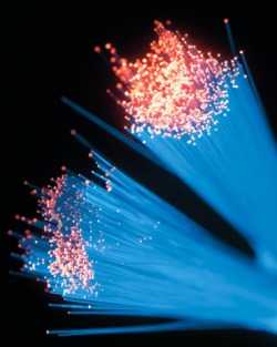
One possibility that biophotonics presents for solving this problem is the use of fiber optics to enable doctors to bring their tools into the patient rather than removing a part of the patient. The function of fiber optic cables is basically to move light from one place to another. They have total internal reflection, which means that the light that goes in on one side of the cable comes back out on the other side with very minimal distortion. So a possible tool for the future would be inserting an electro-optical probe into the patient to bring light-related information (such as a Raman spectrum), to the tools that help analyze the material that would otherwise have to be removed from the patient. A great deal of work and research remains to be done to make this an actual reality.
A final interesting bit of information. An interesting aspect of our learning has been hearing about the many obstacles that stop potential cures from getting used in hospitals. Many research projects are abandoned due to lack of investor capital. The U.S. spends far more on research than other countries (which contributes to our high healthcare costs), but even so, research has to ultimately take on the form of a product to be profitable. If research lacks a vision for how it will be brought to the marketplace in a profitable fashion, the odds are that it won’t survive.
I’ve really enjoyed learning about biophotonics this quarter. I’m still ultimately convinced of the direction I’m heading, but the class has reinforced my feeling that I could be happy doing a myriad of things with my life. There is so much to learn and discover in the world that it’s hard to choose what to focus on.
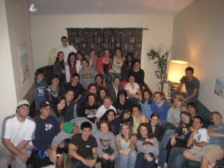
Forty-five people...one house...no showers allowed.
On a more personal note, the quarter has continued to be busy. This last weekend, I attended another retreat (no planning for me this time!), this time with the freshmen of College Life, the fellowship where I think I feel most at home. It was quite a weekend with forty-five people crammed into a beach house in Santa Cruz. On the last night of the retreat, there was a time where all forty-five people gave a short testimony of where they’d come from and how they felt about their current relationship with God.
I think it was the deepest group experience I’ve ever encountered. While some people gave testimonies of relatively peaceful lives, others shared stories of broken pasts, the likes of which I’ve never heard personally. There were stories of molestation, suicidal friends, deaths in the immediate family, and overwhelming loneliness. The same stories were filled with joy, hope, freedom, and forgiveness. After this, the thirteen guys at the retreat met together and talked and prayed for four hours. It was truly amazing.
And we’re only five months in.
 RSS Feed
